- What are the false positive and negative rates for the scanner in comparison with the final histology results?
- GS – We generated 60 images of prostates and had 1 false positive and 1 false negative.
- The Histolog Scanner is capable of offering information about the apical margin which NeuroSAFE is not, do you agree?
- GS – I think this is an interesting area for further work. In theory yes, but in order to be useful in this way a controlled secondary resection must be feasible.
- AM – Neurosafe is not appropriate to evaluate margins at apex level. As Histolog Scanner examines the morphology of a certain surface area, we could potentially use “preferred areas” to examine based on preoperative findings of the tumour.
- Histopathology utilizes a set of features like nucleolar size or absence of basal cells to discriminate between benign and cancerous glands. Does your learning curve contain a set of differential diagnosis features for the same purpose?
- AH – As the rate of positive margins routinely was low, we modified and created a technique to expose prostatic tissue to learn and increase the exposure to images of both benign and cancerous glands. This has increased the learning curve to recognise the conglomerate of nuclear and cytoarchitectural features to characterise the glands.
- How much operating table time do you think will be saved by utilizing this platform to make the resect decision when compared to standard practice/NeuroSAFE?
- GS – This is something that needs more formal evaluation. Generally, we find that image acquisition takes 3 or 4 minutes. The pathologist can analyse the images in a similar time. So, we are talking about 10 minutes overall. When we do NeuroSAFE at my center where geography is a bit of an issue and we have to get the specimen to the lab to get it processed it takes us about 45-55 minutes to get an answer from NeuroSAFE.
- AM – I have very little experience with NeuroSAFE. As we did not lay this out logistically in our department it took us far too long, an hour or so. It takes not even minutes to have the morphological data with the Histolog scanner. It keeps operative times low.
- Do you also view resected tissue using the Histolog or does that follow standard processing?
- AM – Yes, we do that to understand the difference between tumour morphology, benign morphology, and non-prostate morphology. Nowadays when they take definitive morphology they work with slices. They don’t know about the whole surface. So, I foresee that they will take the morphology of the whole surface to see where they need to cut or use that information to add to their definitive pathology result.
- GS – So, the morphology of the secondary resected bundle is really interesting. In our study on NeuroSAFE, we find tumors in the secondary resection where we have a positive margin in about 40% of cases. When they use NeuroSAFE in the Martini-Klinik where it is about 50%. This follows on from the point I made in the discussion about only being able to look at what you have taken out, not what you have left behind. Also, it may allude to what Alex mentioned about processing errors. If we could find a way to orientate the bundle as it comes out, you could perhaps do an en face and perhaps get a more accurate assessment of whether there is tumour there or not.
- The main problem I see is related to the prostate surface, which is rounded. So, especially for large prostates, how can you avoid artifacts related to lack of contact between the prostate surface and the confocal microscope?
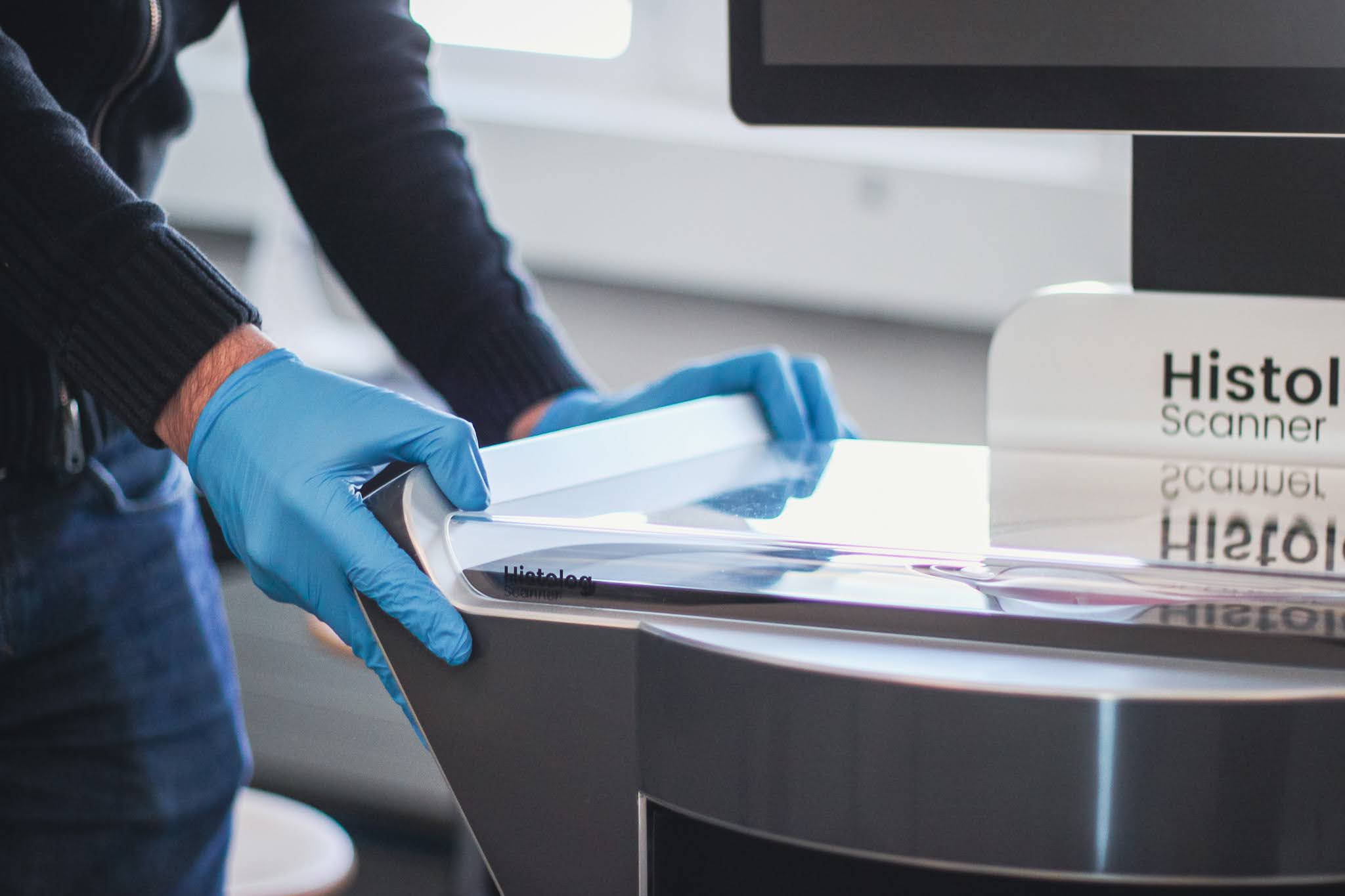
- GS – With our limited experience of 60 or so cases we have imaged prostates up to 150 grams. You occasionally need to compress the specimen onto the objective lens. However, the objective lens is big, being 17 cm2 wide on the histology scanner. So, you can image the whole surface provided you can press the specimen down. We have malleable weights that we use. We also have a device that uses elastic bands to hold it in the right position.
- AM – Large prostates are not a problem when using the Histolog scanner. When it is a big surface it might be the case that you have to take 2 images from one side.
- What kind of histological alteration led to the false positive result? Adenosis? Was a pathologist involved?
- AH – The crowding of glands in adenosis/ atrophy was regarded as malignant by a junior pathologist. Once the conglomerate (both nuclear and cytoarchitectural) of features were taken into account a correct diagnosis was made. Like any other image analysis including slide microscopy; the learning curve will be dependent on exposure and experience of the number of cases.
- Is it possible to scan a second margin after prostate removal using the Histolog scanner?
- GS – Yes, in theory.
- What is your management in the case of a benign gland on a histology margin?
- GS- I think that resection of an entire half of a man’s erectile nerves on the basis of concern regarding him having a detectable PSA, but not cancer recurrence, is excessive, to say the least. I do not do a secondary resection for benign glands at the surgical margin.
- AM – I have a slightly different view on this. In case of a significant margin (>3mm) of benign prostatic tissue, I would re-resect the tissue in that region. In my opinion, oncological outcome exceeds functional outcome (at least potency)
- Given that final pathology is performed with transverse slices, how do you match margin locations to your en face Histolog images?
- AM – I don’t see any reason why to do so. In final pathology, you do slices every 3 mm. I could imagine that Histolog becomes part of the protocol of the definitive pathology report to define better positive margin status and surface of positive margin area.
- These benign glands seem to have “prominent nucleoi”, was IHC later performed to rule out tumor?
- AH- The diagnosis of prostate cancer is made on a conglomerate of multiple nuclear and cytoarchitectural features and just one features would not prompt a diagnosis. The glands present ones at the inked margin did not have nucleoli and none of the other features were an indicator of malignancy. There can be surrounding cancer but as long as these are not at the margin it would not be called a positive margin. Therefore immunohistochemistry was not done.
- Since the image resolution is lower than conventional H&E images, do you think this is enough to say whether there are cancer cells or not on the margin? Would you be so confident to make any statement on margin status just based on CFM and decide your surgical strategy afterward?
- GS – When there are no glands at the margin this is straight forward and a call can be made based on CFM images. Where glands are present on the surface, the question of whether they can be identified as benign of cancerous is important, the initial signs are promising but more work is required.
- A question regarding health economics: how much does a Histolog scanner cost Will the potential benefits of using Histolog scanner cover this?
- GS – The initial cost is relatively high but minimal consumables and training costs and the possibility of use in other tissue types means that the costs are likely to be covered in high volume surgical centres.
Speakers
Alexandre Mottrie MD PhD
Head of the Urological Department OLV Hospital, Aalst, Belgium & CEO of ORSI Academy, Ghent, Belgium
Prof. Dr. Alexandre Mottrie is a pioneering figure in urological oncology and robotic surgery. Graduating in 1988 from the Catholic University of Leuven, Belgium, he completed his residency at the Johannes Gutenberg University of Mainz, Germany, in 1994. Since 1996, he’s been a urologist in Belgium, developing significant expertise in robotic surgery. In 2010, he founded ORSI Academy, a training center for robotic and minimal-invasive surgery. Dr. Mottrie is renowned for his extensive scientific contributions, including organizing international congresses and masterclasses. He’s received multiple awards, including the “Golden Telescope Award” and the “Saint-Pauls’ medal.”
Greg Shaw MD FRCS (Urol)
Lead for Robotic Urology, UCLH
Professor of Urology, UCL
Professor Greg Shaw is a distinguished Consultant Urological Surgeon based in London, UK. He graduated from St Bartholomew’s Medical College in 1999. His career has been marked by a profound interest in prostate cancer, developed during his MD research at St Bartholomew’s Hospital and furthered during a Lectureship at The University of Cambridge. Here, he contributed significantly to prostate cancer research. Prof. Shaw completed his specialist training in North London, also earning a Fellowship from the Royal College of Surgeons of England. His expertise includes robotic surgery, and he is committed to applying the latest scientific knowledge in prostate cancer diagnosis and treatment.
Dr Aiman Haider, MD, FRCPath
Consultant histopathologist (Urological Pathology) Department of Cellular Pathology, UCLH, London
Dr. Aiman Haider is a distinguished Consultant Histopathologist specializing in Urological Pathology at the Department of Cellular Pathology, UCLH, London. She serves as the Meetings and Education Secretary for the British Association of Urological Pathologists and holds an Honorary Lecturer position at UCL London. Dr. Haider plays a crucial role in several influential committees, including the EAU/ESU Working Group on Imaging Focal Uropathology and the Genitourinary Pathology Society (GUPS). Her dedication to advancing the field is evident through her significant research contributions, active involvement in the HSL/UCL Research Innovations Committee, and participation in the prestigious Marcel Levi Uropathology Fellowship. Dr. Haider’s expertise significantly enhances clinical surgical trials and translational research in pathology.


